Quantitative analysis of blood velocity in the retina.
The goal of het project was to develop a new method to measure blood flow velocity in small blood vessels in het retina, in collaboration with Heidelberg Engineering, a leading manufacturer of opthalmic imaging devices. Blood flow velocity is an important indicator of retinal health.
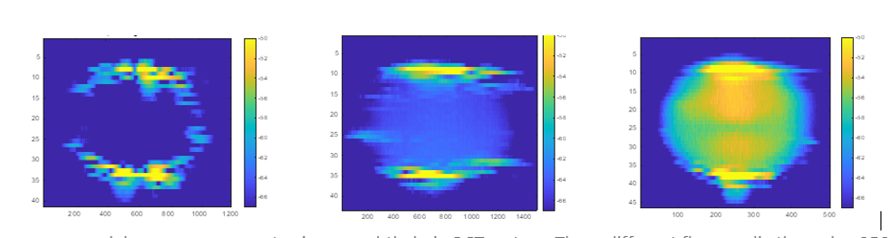
This project determined the best method to determine blood flow velocity by a method called Phasor Pair decorrelation, where the decorrelation is determined between two electromagnetic fields (lightwaves) that are reflected from the retina after consequtive measurements. The precision or statistical fluctuation of the quantitative velocity estimation depends on the number of repeated measurements and the method to determine quantitative flow velocity. Both a method to determine quantitative flow velocity and a model for the prediction of the statistical fluctuations of velocity estimations was developed to analyse and optimise the estimation precision. The method and model were validated by phantom measurements in a bulk scattering medium as well as in intralipid solution in a capillary. Based on the model the number of repeated measurements to achieve a certain velocimetry precision was predicted. To help translate these results on intralipid flow towards in-vivo application, they have constructed a flow system for live human blood flowing through retina-mimicking silicone elastomer phantoms. This is required for the experimental realisation of real blood flow measurements, and a verification of the extension of their theory for point scatterers to real blood. One important result from the blood flow system is the reproduction of the famous ‘hourglass’ figure, seen on in-vivo retina OCT scans. This is only seen with blood as a flow medium, and points to a good reproduction of in-vivo conditions in their system.
In conclusion, both a method to determine quantitative flow velocity and a model for the prediction of the statistical fluctuations of velocity estimations was developed to analyse and optimise the estimation precision for phase based velocimetry methods. The theory was verified in a flow system with intralipid (point scatterers).
To verify the theory for human blood, a system was developed to flow living human blood through small capillaries, while controlling temperature, flow velocity, and oxygenation level. Human blood flow can be controlled and flowed outside the body, in a way that is optically accessible to OCT and SLO systems which mimicks retinal vasculature. Experiments with this system to verify the flow velocity theory on human blood are in progress.