The use of light and magnetism to reveal stem cell-based repair of hearing
It is estimated that 5% of people worldwide suffer from disabling hearing loss in both ears. The most common type of hearing loss is caused by permanent damage to the cochlea and this cannot be medically treated. The only widely successful therapy for patients who are profoundly deaf or severely hearing-impaired is a cochlear implant (CI). So far, more than 250,000 patients have been implanted worldwide. A CI is a small, complex electronic device, which stimulates the auditory nerve providing functional hearing to most recipients. However, there exists a large variability in CI-performance. This may be due to differences between patients in the degree of auditory neuron loss.
Stem-cell-based therapy has the potential to be used to repair auditory neurons and thereby improve CI efficacy. To investigate this hypothesis, it is required that stem cells, after transplantation into the cochlea of a deaf animal, are visualized for a long period of time. Different visualization techniques, such as bioluminescence, fluorescence and magnetic resonance imaging (BLI, FLI and MRI), allow tracing of stem cells in the living test animal and post-mortem. The combination of these techniques is possible because the stem cells are genetically manipulated to give light (both bioluminescent and fluorescent light) and subsequently loaded with magnetic nanoparticles to allow also MRI visualization. Furthermore, it provides indispensable information about migration, survival and regenerative capacity of transplanted stem cells. However, visualization of stem cells in the inner ear of living animals has never been achieved and requires proper post-mortem validation using high-resolution microscopy techniques.
The purpose of this study is to corroborate information gained from BLI and MRI in the living animal and afterwards, post-mortem, by fluorescence high-resolution microscopy. Additional aims are: (1) to determine the environmental adaptation of the cells; (2) to understand the mechanism of recovery and (3) to determine if any adverse effect followed implantation. To accomplish this, the following experiment is organised: stem cells derived from mouse hair follicles will be implanted into the cochlea of deafened guinea pigs and migration and survival will be monitored in the living animal by means of BLI and MRI for a period of 4 months. Post-mortem high-resolution fluorescence microscopy will be performed to detect the stem cells and to determine their functional effect. The complimentary information from all techniques will illustrate the 4-month history of the stem cells. This information is needed to understand the process of stem-cell-based therapy of the auditory nerve. This may lead to further improvement of hearing in CI recipients. MED-EL is dedicated to novel research in this field and has been supporting this project for years.
For more information, see this website.

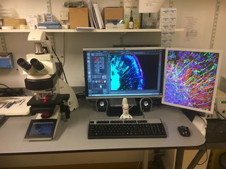
Fig 1: This image shows the microscope, equipped with a near-infra red module, acquired with the TKI-LSH Match funding. The left screen shows a picture of mouse hair inner ear tissue stained for different types of neuronal cells . The right screen shows hair follicle stem cells after outgrowth from the hair follicle.


