Novel ECG algorithms to better understand and visualize the underlying pathophysiology of STT-segment changes (STT-vision) during disease development and progression in patients with genetic heart diseases
Novel ECG algorithms to better understand disease development and progression in inherited cardiomyopathies
The STT vision project is a collaboration between the University Medical Center Utrecht, Radboud University Medical Center and ECG Excellence, a Dutch startup with the ambition to change the way we use the ECG to detect heart diseases. Within this project we will use combinations of 67 lead ECG recordings, 12 lead ECG recordings, newly developed ECG interpretation algorithms (CineECG and ECG imaging) and a 3D visual guided electrode placement technology.
The described ECG techniques will be used in the STT-vision project to improve our understanding of disease onset, disease development and disease progression in patients with genetic heart diseases. This is of great importance because we want to identify individuals at risk for life threatening ventricular arrhythmias. Using novel ECG techniques we aim to unravel and better understand the underlying electrical changes during the recovery phase of the heart. This will aid earlier and more effective treatment of the disease and will reduce stress and uncertainties both in patients and clinicians. Our ECG method also provides a simple, fast and non-invasive monitoring solution.
In the STT-vision project, we first optimized our novel ECG technique - a computerized heart model – to model the recovery phase of the heart in healthy individuals. We then validated this technique against the gold standard (invasive data) in patients with (genetic) heart diseases, observing strong agreement with our model. Next, we applied the computerized heart model to a cohort of 140 patients with a genetic heart disease at risk of cardiac arrest. We are now evaluating its ability to detect early disease onset, disease development, and disease progression in these patients. The first results are promising as distinct differences can be observed between patients and control subjects (Figure). Distinct differences were also observed using our CineECG technique, even in patients showing a normal 12-lead ECG.
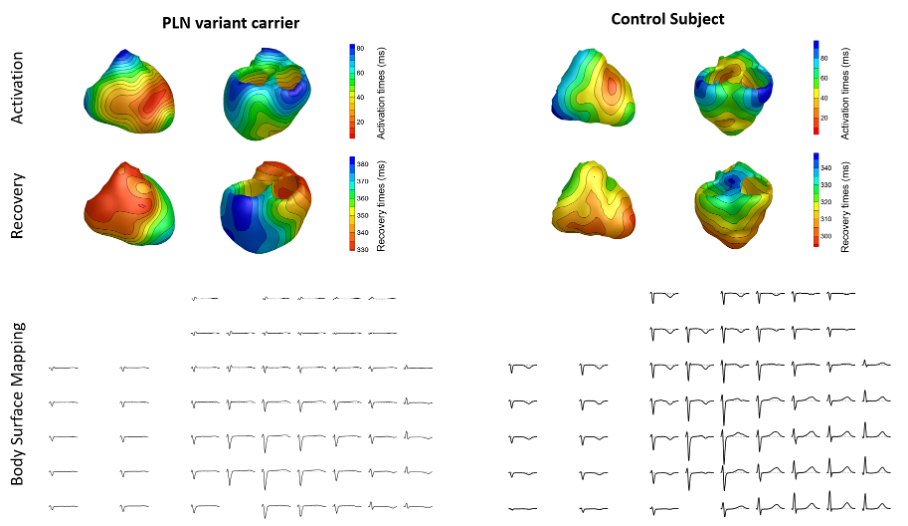
Figure Examples of the estimated activation/depolarization sequence (upper row), recovery/repolarization sequence (middle row), and the corresponding body surface maps (lower row) in a PLN carrier (left column) and a control subject (right column). The areas of early activation and recovery are shown in red, areas of late activation and recovery times are shown in blue. The activation sequence in the PLN-carrier shows isochronal crowding when compared to the control subject, the recovery sequence shows an abnormal order of repolarization where the apex and inferoposterior wall show late repolarization.
Andere projecten
In elk PPS-project werken kennisinstellingen, bedrijven en zorgpartijen samen aan een concrete gezondheidsvraag. Bekijk het volledige projectoverzicht.
Bekijk de programma’s
Binnen onze programma’s werken bedrijven, kennisinstellingen en regionale partners samen aan innovaties die bijdragen aan betere gezondheid en zorg.
Bekijk de partnerschappen
Strategische publiek-private partnerschappen (PPP's) zijn meerjarige samenwerkingen tussen bedrijven, kennisinstellingen en andere partijen.
Hoe kunnen we je helpen?
Heb je een vraag of wil je meer weten over Health Holland? We brengen je graag in contact met de juiste mensen, kennis en mogelijkheden om gezondheidsinnovatie te versnellen.
We hebben uw bericht ontvangen
We nemen binnen 3 tot 4 werkdagen contact met je op. Is je vraag dringend? Neem dan gerust direct contact met ons op. Hieronder zie je de contactoptie die het beste past bij je onderwerp.
Opnieuw beginnen
Er ging iets mis bij het versturen van het formulier. Probeer het opnieuw.