Light tissue interactions in ophthalmology
Despite enormous improvement in ophthalmic imaging techniques over the last few decades, the information that can be gained from light-tissue interactions is still not fully exploited. New microscopic and interferometric techniques are still being developed that provide better resolution based on new contrast mechanisms. The project is a collaboration between the VU University Department of Physics, the Amsterdam UMC department of Ophthalmology, and Heidelberg Engineering, a leading European manufacturer of ophthalmic diagnostic devices.
The result of this research will lead to new methods to characterise a variety of phthalmic diseases, such as glaucoma, age related macular degeneration and diabetic retinopathy which are leading causes of blindness in the developed world.
In this project the consortium will investigate how tissue properties are encoded on light signals reflected from retinal layers in the eye. In particular they will investigate how motion (blood flow) can be detected by analysing the probability distribution of phasors due to moving scatterers, how the polarisation state of light can be used to determine the presence of fibrotic structures, how the attenuation coefficient of tissue can be determined in a reliable manner and if it can be used to determine axon density in the retinal nerve fiber layer.
The consortium has developed a new method to quantify flow in the retina and verified the method in phantoms, they have developed a new method to analyse the polarisation state reflected from the retina and visualised fibrotic tissue in AMD patients, and they have for the first time determined the scleral collagen structure in vivo.
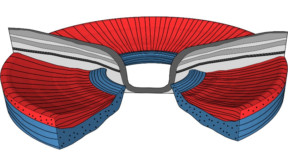
Model of peripapillary scleral orientation. A radially oriented fiber layer (red) precedes a circumferentially orientated fiber layer (blue). Close to the ONH, the circumferentially oriented layer displaces the radially oriented fiber layer. Copyright Translational Vision Science & Technology October 2020, Vol.9, 21. doi:https://doi.org/10.1167/tvst.9.11.21